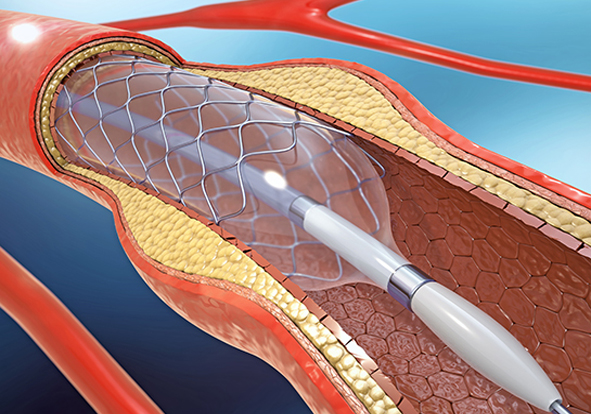
Angiography, also known as arteriography, is an imaging technique which is used to look inside the lumen of the heart, arteries, veins and different blood vessels. This test is used to study if there is any blocked, narrowed, enlarged, or malformation within the arteries or veins in any part of the body like heart, brain, legs, abdomen etc.
Angioplasty is usually combined with stent placement. After the fatty plaque is pushed out and the blocked vessel is opened, a wire-mesh tube (stent) is then placed in this area. The stent is left in the artery permanently to help keep the artery open (scaffolding the vessel). With Angioplasty alone - without stent placement re-narrowing of an artery (restenosis) happens in about 30-40 percent of cases therefore stent placement is widely recommended.
Why is it done?
Angioplasty is done to treat arterial blockages. The blockages mainly occur due to:
- Atherosclerosis: The slow buildup of plaques in your blood vessels.
- Coronary artery diseases: The blockage of the arteries carrying oxygenated blood to heart muscles.
- Peripheral artery diseases: .The narrowing of arteries in legs and arms.
- Carotid artery stenosis: The narrowing of neck arteries supplying blood to brain.
- Renal vascular hypertension: Where high blood pressure is caused by the narrowing of arteries in the kidney.
- Angina: Severe chest pain or tightness in the chest area felt on the middle to left side of the chest.
- Narrowing of arteries in chest, abdomen, pelvis, arms, and legs.
- Feeling short of breath
- Heart attack.
- In case the dialysis fistula and grafts are narrowed or blocked.
Symptoms
Angioplasty is done to treat arterial blockages. The blockages mainly occur due to
- Atherosclerosis: The slow buildup of plaques in your blood vessels.
- Coronary artery diseases: The blockage of the arteries carrying oxygenated blood to heart muscles.
- Peripheral artery diseases: The narrowing of arteries in legs and arms.
- Carotid artery stenosis: The narrowing of neck arteries supplying blood to brain.
- Narrowing of arteries in chest, abdomen, pelvis, arms, and legs.
- Renal vascular hypertension: Where high blood pressure is caused by the narrowing of arteries in the kidney.
- In case the dialysis fistula and grafts are narrowed or blocked.
- Angina: Severe chest pain or tightness in the chest area felt on the middle to left side of the chest.
- Feeling short of breath.
- Heart attack.
However, it should be noted that angioplasty isn’t a treatment for all types of blockages. It is not done in case of:
in case you had an heart attack, the recovery period will be higherin case you had an heart attack, the recovery period will be higher
- The main artery that brings blood to the left chambers of your heart is narrowed.
- If your heart muscles are weaker.
- In case you have multiple numbers of diseased blood vessels.
- In case you are diabetic, angioplasty can be carried out but with additional precaution.
- In a nutshell, the decision of angioplasty is taken by the doctors depending on the extent of your heart damage and your overall medical condition.
Diagnosis
Generally the most common sign a patient experiences is angina. Depending on the nature of angina, the doctors performs cardiac tests like ECG, stress test, along with additional chest X-Ray, chest CT scan, angiogram, cardiac MRI etc.
Post Operative Care<
- In case you had a non-emergency procedure, you will be released after 24 hours observation.
- In general you can return to your normal routine within a week of procedure
- in case you had an heart attack, the recovery period will be higher
- Drink plenty of fluids to flush the contrast material out of system
- Avoid any kind of strenuous activity or any kind of heavy-lifting
Pacemaker Implantation
A pacemaker is a small device that's placed in the chest or abdomen to help control abnormal heart rhythms. This device uses electrical pulses to prompt the heart to beat at a normal rate. Pacemakers are used to treat arrhythmias (ah-RITH-me-ahs). Arrhythmias are problems with the rate or rhythm of the heartbeat.
A Biventricular Pacemaker is a pacemaker that paces on both sides of the heart. In selected cases, resynchronising the heart in this fashion can improve cardiac function.
The pacemaker consists of two components, the generator, or battery, and the leads or wires. The generator is about the size of two stacked 50c coins and is implanted under the skin beneath your collarbone. This is connected to the leads or wires which rest inside your heart. The wire is very soft and flexible and can withstand the twisting and bending caused by body movements.
Types
The type of pacemaker that you may need depends solely on the condition of your heart. After a detailed diagnostic evaluation and various test your doctors will decide the type of pacemaker right for your heart. Normally there are three different kind of pacemakers that are used
- Single chambered pacemaker: This is the most commonly used pacemaker type.
- Dual chamber pacemaker: Here two lead wires are taken which are connected to both the right atrium and right ventricle to facilitate simultaneous relaxation and contraction of heart in a proper rhythm.
- Bi ventricular pacemaker : This is also known as CRT for cardiac resynchronization therapy. Implantation of pacemaker is an invasive surgery. And thus before the surgery your doctors will prepare you for a couple of tests to get a full overview of your heart conditions. These tests includ
- An electrocardiogram to get a clue about the nature of irregular heartbeats you are having
- Holter monitoring or ambulatory monitoring which will record your heart rate for an entire period of 24 hours during which time you will be asked to keep a record of all the activities you were doing
- An echocardiogram get images of your heart condition and to see the thickness of your heart muscles
- Stress test along with an echocardiogram or nuclear imaging to record the oxygen consumption levels by your heart


